


Hot Products
YSX500D 50kW DR system set up and put into service in Cambodia.
YSENMED YSX500D 50kW digital x-ray system has been successfully set up and put into service in a hospital in Cambodia.
YSX056-PE serving as a vehicle-mounted x-ray in the Philippines
YSX056-PE 5.6kW portable x-ray unit has been adapted to fit on a truck, to provide mobile x-ray examination service for remote communities in the Philippines.
X Ray Machine To Zimbabwe
x ray machine, 50KW x ray machine
Microscope To Malawi
Achromatic objectives: 4X、10X、40X(S), 100X(S、Oil) Wide field eyepiece: WF10X(WF16X for option) Eyepiece head: Sliding binocular head inclined at 45° Stage: Double layer mechanical stage size 140X140mm, moving range 75X45mm Focusing: Coaxial coarse and
Seeing the Unseen: The Power of Optical Coherence Tomography in Eye Care
Views : 6398
Update time : 2024-05-28 11:05:38
1. Introduction
In the realm of eye care, the ability to see what lies beneath the surface has long been a challenge. However, with the advent of Optical Coherence Tomography (OCT), that challenge has been met with groundbreaking technology that allows for the visualization of intricate eye structures in unprecedented detail. In this article, we embark on a journey through the world of OCT, exploring its mechanisms, applications, and the profound impact it's having on the diagnosis and treatment of various eye conditions.
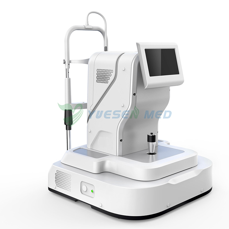
2. Understanding Optical Coherence Tomography (OCT)
Optical Coherence Tomography, commonly referred to as OCT, is a non-invasive imaging technique that revolutionized the field of ophthalmology. Unlike traditional imaging methods such as ultrasound or MRI, OCT utilizes light waves to create detailed cross-sectional images of the eye's interior. By measuring the reflections of these light waves, OCT generates high-resolution, three-dimensional images of the retina, optic nerve, and other ocular structures.
3. How OCT Works its Magic
To grasp the inner workings of OCT, imagine a beam of light traveling through the eye much like a flashlight piercing through fog. As the light encounters different structures within the eye, such as the retina or the cornea, it reflects back, producing interference patterns. By analyzing these patterns, OCT constructs detailed images with micron-level resolution, allowing clinicians to visualize subtle changes in tissue morphology and identify abnormalities with remarkable precision.
4. Applications of OCT in Eye Care
The versatility of OCT extends across a wide spectrum of eye conditions, making it an indispensable tool in modern ophthalmic practice. From diagnosing and monitoring diseases such as glaucoma, macular degeneration, and diabetic retinopathy to guiding surgical interventions like refractive surgery and retinal detachment repair, OCT plays a pivotal role in the management of ocular health. Its ability to detect early signs of pathology enables timely intervention, leading to improved outcomes and preservation of vision.
5. Advantages of OCT Over Traditional Methods
Compared to conventional imaging modalities, OCT offers several distinct advantages. Unlike fluorescein angiography or fundus photography, OCT is non-invasive, painless, and does not require the injection of contrast agents. Moreover, OCT provides real-time, high-resolution images that offer unparalleled insights into the structural integrity of the eye. Its ability to visualize microstructural changes allows for early detection of disease progression and facilitates personalized treatment strategies.
6. Making a Difference in Patient Care
The impact of OCT on patient care cannot be overstated. By providing clinicians with detailed anatomical information in a matter of seconds, OCT empowers them to make informed decisions regarding patient management. Whether it's monitoring disease progression, assessing treatment efficacy, or guiding surgical planning, OCT enhances the quality of care delivered to patients, ultimately improving outcomes and enhancing their quality of life.
7. The Future of OCT: Innovations and Beyond
As technology continues to evolve, so too does the field of OCT. Recent advancements, such as swept-source OCT and adaptive optics, have expanded the capabilities of OCT, enabling faster imaging speeds, deeper tissue penetration, and enhanced visualization of microscopic structures. Furthermore, ongoing research efforts aim to integrate OCT with artificial intelligence algorithms for automated image analysis and diagnosis, paving the way for more efficient and accurate patient care.
8. Overcoming Challenges and Limitations
Despite its numerous benefits, OCT is not without its challenges. Cost can be a significant barrier to widespread adoption, particularly in resource-limited settings. Additionally, interpretation of OCT images requires specialized training, and certain eye conditions, such as cataracts or severe corneal scarring, can affect image quality, limiting the utility of OCT in these cases. However, ongoing efforts to improve accessibility, affordability, and image interpretation are underway to address these challenges and expand the reach of OCT to underserved populations.
9. Conclusion
In conclusion, Optical Coherence Tomography (OCT) represents a paradigm shift in the field of eye care, offering clinicians unprecedented insights into the structure and function of the eye. From its ability to detect subtle changes in tissue morphology to its role in guiding surgical interventions, OCT has transformed the way we diagnose, monitor, and treat ocular diseases. As we look to the future, the continued evolution of OCT promises to further enhance our understanding of the eye and revolutionize the way we deliver care to patients worldwide.
FAQs:
What is Optical Coherence Tomography (OCT)?
Optical Coherence Tomography, or OCT, is a non-invasive imaging technique used in ophthalmology to produce high-resolution, cross-sectional images of the eye's interior. It utilizes light waves to create detailed images of ocular structures, including the retina, optic nerve, and cornea.
How does OCT differ from other imaging methods like MRI or ultrasound?
Unlike MRI or ultrasound, which rely on different physical principles for imaging, OCT uses light waves to create images of the eye's interior. This allows for higher resolution and more detailed visualization of ocular structures, making OCT particularly well-suited for diagnosing and monitoring eye conditions.
What are the common applications of OCT in eye care?
OCT has a wide range of applications in eye care, including the diagnosis and monitoring of conditions such as glaucoma, macular degeneration, diabetic retinopathy, and retinal detachment. It is also used in refractive surgery to assess corneal thickness and shape, as well as in the management of optic nerve disorders.
Is OCT safe for patients?
Yes, OCT is considered safe for patients. It is a non-invasive imaging technique that does not involve the use of ionizing radiation or contrast agents. However, as with any medical procedure, there may be rare instances of adverse reactions or discomfort, but these are minimal and usually transient.
Can anyone undergo OCT imaging?
While OCT is safe for most patients, there are certain factors that may affect its utility. For example, patients with severe corneal scarring or opacities may have poor image quality, limiting the diagnostic accuracy of OCT. Additionally, OCT may not be suitable for patients who are unable to sit still or maintain fixation during the imaging process. However, in general, OCT is a valuable tool that can benefit a wide range of patients in the diagnosis and management of various eye conditions.
In the realm of eye care, the ability to see what lies beneath the surface has long been a challenge. However, with the advent of Optical Coherence Tomography (OCT), that challenge has been met with groundbreaking technology that allows for the visualization of intricate eye structures in unprecedented detail. In this article, we embark on a journey through the world of OCT, exploring its mechanisms, applications, and the profound impact it's having on the diagnosis and treatment of various eye conditions.
2. Understanding Optical Coherence Tomography (OCT)
Optical Coherence Tomography, commonly referred to as OCT, is a non-invasive imaging technique that revolutionized the field of ophthalmology. Unlike traditional imaging methods such as ultrasound or MRI, OCT utilizes light waves to create detailed cross-sectional images of the eye's interior. By measuring the reflections of these light waves, OCT generates high-resolution, three-dimensional images of the retina, optic nerve, and other ocular structures.
3. How OCT Works its Magic
To grasp the inner workings of OCT, imagine a beam of light traveling through the eye much like a flashlight piercing through fog. As the light encounters different structures within the eye, such as the retina or the cornea, it reflects back, producing interference patterns. By analyzing these patterns, OCT constructs detailed images with micron-level resolution, allowing clinicians to visualize subtle changes in tissue morphology and identify abnormalities with remarkable precision.
4. Applications of OCT in Eye Care
The versatility of OCT extends across a wide spectrum of eye conditions, making it an indispensable tool in modern ophthalmic practice. From diagnosing and monitoring diseases such as glaucoma, macular degeneration, and diabetic retinopathy to guiding surgical interventions like refractive surgery and retinal detachment repair, OCT plays a pivotal role in the management of ocular health. Its ability to detect early signs of pathology enables timely intervention, leading to improved outcomes and preservation of vision.
5. Advantages of OCT Over Traditional Methods
Compared to conventional imaging modalities, OCT offers several distinct advantages. Unlike fluorescein angiography or fundus photography, OCT is non-invasive, painless, and does not require the injection of contrast agents. Moreover, OCT provides real-time, high-resolution images that offer unparalleled insights into the structural integrity of the eye. Its ability to visualize microstructural changes allows for early detection of disease progression and facilitates personalized treatment strategies.
6. Making a Difference in Patient Care
The impact of OCT on patient care cannot be overstated. By providing clinicians with detailed anatomical information in a matter of seconds, OCT empowers them to make informed decisions regarding patient management. Whether it's monitoring disease progression, assessing treatment efficacy, or guiding surgical planning, OCT enhances the quality of care delivered to patients, ultimately improving outcomes and enhancing their quality of life.
7. The Future of OCT: Innovations and Beyond
As technology continues to evolve, so too does the field of OCT. Recent advancements, such as swept-source OCT and adaptive optics, have expanded the capabilities of OCT, enabling faster imaging speeds, deeper tissue penetration, and enhanced visualization of microscopic structures. Furthermore, ongoing research efforts aim to integrate OCT with artificial intelligence algorithms for automated image analysis and diagnosis, paving the way for more efficient and accurate patient care.
8. Overcoming Challenges and Limitations
Despite its numerous benefits, OCT is not without its challenges. Cost can be a significant barrier to widespread adoption, particularly in resource-limited settings. Additionally, interpretation of OCT images requires specialized training, and certain eye conditions, such as cataracts or severe corneal scarring, can affect image quality, limiting the utility of OCT in these cases. However, ongoing efforts to improve accessibility, affordability, and image interpretation are underway to address these challenges and expand the reach of OCT to underserved populations.
9. Conclusion
In conclusion, Optical Coherence Tomography (OCT) represents a paradigm shift in the field of eye care, offering clinicians unprecedented insights into the structure and function of the eye. From its ability to detect subtle changes in tissue morphology to its role in guiding surgical interventions, OCT has transformed the way we diagnose, monitor, and treat ocular diseases. As we look to the future, the continued evolution of OCT promises to further enhance our understanding of the eye and revolutionize the way we deliver care to patients worldwide.
FAQs:
What is Optical Coherence Tomography (OCT)?
Optical Coherence Tomography, or OCT, is a non-invasive imaging technique used in ophthalmology to produce high-resolution, cross-sectional images of the eye's interior. It utilizes light waves to create detailed images of ocular structures, including the retina, optic nerve, and cornea.
How does OCT differ from other imaging methods like MRI or ultrasound?
Unlike MRI or ultrasound, which rely on different physical principles for imaging, OCT uses light waves to create images of the eye's interior. This allows for higher resolution and more detailed visualization of ocular structures, making OCT particularly well-suited for diagnosing and monitoring eye conditions.
What are the common applications of OCT in eye care?
OCT has a wide range of applications in eye care, including the diagnosis and monitoring of conditions such as glaucoma, macular degeneration, diabetic retinopathy, and retinal detachment. It is also used in refractive surgery to assess corneal thickness and shape, as well as in the management of optic nerve disorders.
Is OCT safe for patients?
Yes, OCT is considered safe for patients. It is a non-invasive imaging technique that does not involve the use of ionizing radiation or contrast agents. However, as with any medical procedure, there may be rare instances of adverse reactions or discomfort, but these are minimal and usually transient.
Can anyone undergo OCT imaging?
While OCT is safe for most patients, there are certain factors that may affect its utility. For example, patients with severe corneal scarring or opacities may have poor image quality, limiting the diagnostic accuracy of OCT. Additionally, OCT may not be suitable for patients who are unable to sit still or maintain fixation during the imaging process. However, in general, OCT is a valuable tool that can benefit a wide range of patients in the diagnosis and management of various eye conditions.
Related News
Read More >>
 Seychelles Clinic Successfully Equips with YSENMED Ultrasound, Lab & Patient Care Devices
Jul .07.2026
Ribail Babie from Seychelles equipped a private clinic with YSENMED ultrasound, hematology analyzer, biochemistry analyzer, EMG system, patient monitor and more. The customer is highly satisfied with both the equipment performance and our professional ser
 Saudi Distributor Purchases the 5th YSENMED YSX056-PF VET Portable Veterinary DR with Clear Imaging and Reliable Support
Jul .06.2026
A Saudi distributor purchased the fifth YSENMED YSX056-PF VET 5.6kW Veterinary Portable X-ray Unit, praising its sharp image quality, prompt remote support, and expressing plans for future repeat orders.
 Successful Installation of YSOT-YT5D Surgical Table & VG70 Ventilator in Ghana Hospital
Jul .05.2026
YSENMED successfully delivered the YSOT-YT5D 5-Function Electric Surgical Table and VG70 Trolley Turbine Ventilator to a hospital in Ghana. Both units were installed successfully and are now ready to support daily clinical procedures.
 Cameroon Hospital Expands Imaging Capacity with YSENMED 32kW Digital X-Ray Syste
Jul .04.2026
Distributor Hallie reordered the YSENMED 32kW Digital X-Ray Systems for a hospital in Cameroon. The customer praised the excellent image quality and expressed satisfaction with YSENMED's professional after-sales support.
